Neurofibromatosis Tumor Segmentation on Whole-body MRI (WBMRI-NF) Challenge
Datasets Overview
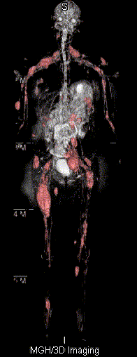
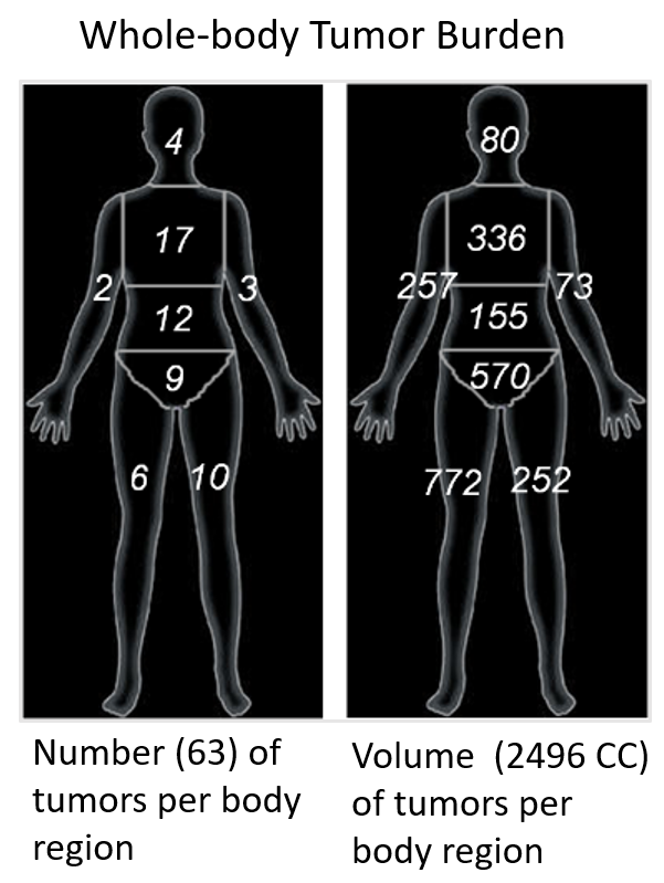
We will provide 400 WBMRI scans (>2400 NF tumors) collected from our clinical working protocol of NF volumetric quantification in WBMRI, each with radiologist-confirmed segmentation masks and volumetric tumor-burden reports. The dataset will be split into 200 for training, 50 for open validation, and 150 for the closed testing.
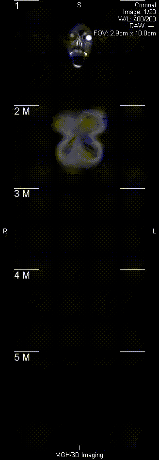
Each WBMRI scan was acquired using a 1.5 Tesla MR scanner with integrated body coil, and no intravenous contrast. Each subject was imaged from head to ankles in the supine position. The entire body was imaged using a coronal fat suppressed fluid sensitive sequence (STIR). The image sizes vary between 320X1000X20 and 400X1200X40. Each scan is saved in a single file (.nii) in NIFTI format, which is converted from de-identified DICOM files.